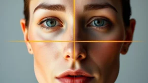
Facial Anatomy Glossary: Anthropometric Landmarks
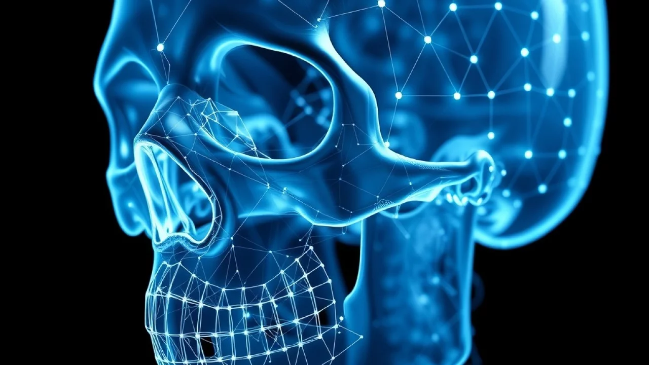
Facial Anatomy Glossary: Anthropometric Landmarks, Craniofacial Terms, and 3D Mesh Nodes
The definitive anatomical reference to cephalometric landmarks, mandibular gonial angles, zygomatic dimensions, palpebral fissures, and 478 3D AI facial nodes.
Whether you are exploring facial plastic surgery, orthodontic bite correction, computer vision landmark engineering, or aesthetic modeling analysis, the scientific study of facial structure relies on a precise anatomical language. Terms like Gonion, Glabella, Menton, Bizygomatic Width, and Palpebral Fissure are not abstract jargon; they are the exact physical coordinates used by surgeons, orthodontists, and computer scientists to map human beauty, developmental health, and facial symmetry.
Historically, human facial geometry was quantified by physical calipers directly touching the skin or measured across lateral cephalometric radiographs. Today, artificial intelligence and neural landmark detectors track up to 478 high-density 3D coordinates in real time directly through camera lenses. Yet, every single machine learning algorithm is rooted in the same foundational principles of craniofacial anthropometry established over a century ago.
This comprehensive Facial Anatomy Glossary serves as your authoritative reference manual. Below, we break down every essential clinical term, explain its physical anatomical position, explore its aesthetic significance, and demonstrate how classical biology translates into cutting-edge 3D biometric computing.
- Anthropometry vs Cephalometrics: Anthropometry focuses on measuring living soft-tissue surfaces, whereas cephalometrics analyzes bony cranial relationships via standardized radiographic projections.
- Skeletal vs Soft-Tissue Landmarks: Skeletal landmarks (e.g., nasion, bony menton) sit directly on skull bones. Soft-tissue landmarks (e.g., subnasale, endocanthion) sit on the skin and mucosal surfaces.
- The Sagittal Midline: The primary vertical reference plane dividing the cranium into left and right hemispheres, establishing the universal baseline for bilateral facial symmetry.
- From Physical Calipers to 478 3D AI Nodes: Modern artificial intelligence transforms 2D camera pixels into dense 3D spatial point clouds across the eyes, lips, jawline, and nose.
Core Scientific Disciplines
What Is Facial Anthropometry?
Facial Anthropometry is the biological and medical science dedicated to the systematic, quantitative measurement of the human face. Derived from the Greek words anthropos (human) and metron (measure), it provides standardized methodologies to measure linear distances, surface curvatures, volume distributions, and angular relationships across cranial structures.
Pioneered extensively by plastic surgeon and researcher Dr. Leslie Farkas in the late 20th century, facial anthropometry replaced subjective, artistic opinions with rigorous, reproducible population norms. Farkas documented standardized norms across diverse age groups, sexes, and ethnicities, establishing baseline ranges for what constitutes typical, balanced facial architecture.
In practical application, anthropometric norms allow clinicians to identify whether a patient’s nose is disproportionately wide, whether their jaw is retracted (retrognathia), or whether their eyes are abnormally close together (hypotelorism) compared to statistically healthy averages.
What Is Anthropometric Facial Analysis?
Anthropometric Facial Analysis is the clinical and diagnostic practice of applying anthropometric principles to evaluate an individual’s facial morphology. Rather than just collecting raw measurements, anthropometric analysis contextualizes those numbers into proportions, aesthetic ratios, and functional assessments.
Clinicians, maxillofacial surgeons, and aesthetic specialists use anthropometric facial analysis across several critical fields:
Designing precise tissue repositioning for cleft lip and palate repair, trauma reconstruction, and orthognathic jaw alignment surgery.
Analyzing dental occlusion relationships, mandibular growth trajectories, and soft-tissue facial changes throughout treatment.
Training neural networks to identify subtle micro-expressions, facial symmetry indexes, and biometric identity verification.
Digital Landmarks & Biometrics
What Is a Facial Landmark?
A Facial Landmark (also known as a craniofacial fiducial point or anatomical anchor) is a biologically or visually distinctive point on the face that maintains a constant, recognizable anatomical meaning across all human individuals. Examples include the tip of the nose (pronasale), the lowest edge of the chin (menton), and the corners of the mouth (cheilion).
In both clinical medicine and computational geometry, facial landmarks serve as universal reference coordinates. Without landmarks, it would be impossible to calculate meaningful distances, compare two different faces, or evaluate changes before and after cosmetic treatments. Landmarks transform a complex organic face into an organized topological map.
What Are 3D Facial Landmarks?
3D Facial Landmarks are digital spatial coordinate points that represent landmark locations in three-dimensional Cartesian space (X, Y, Z):
- X-Coordinate (Transverse Axis): Defines the horizontal position from left to right across the face.
- Y-Coordinate (Sagittal Axis): Defines the vertical position from the crown of the head to the base of the chin.
- Z-Coordinate (Coronal Depth Axis): Defines the anteroposterior depth—how far forward a feature projects (such as the tip of the nose or cheekbones) relative to the back of the skull.
Traditional 2D landmarks captured on photographs only record X and Y coordinates. This limitation causes severe perspective distortion when a subject tilts or turns their head. 3D facial landmarks overcome this flaw by capturing the true volumetric contours of the face, enabling accurate measurements regardless of pose or camera angle.
What Are 478 Facial Landmarks?
The 478 Facial Landmarks refer to the standardized, high-density 3D surface mesh architecture established by state-of-the-art machine learning models, most notably Google MediaPipe Face Mesh. For decades, computer vision relied on the classical dlib 68-point model, which only marked rudimentary boundaries around the eyes, nose, lips, and lower jawline.
By contrast, the 478-point topology represents an exponential leap forward in biometric resolution. It maps 468 dense surface vertices across the entire facial envelope, plus 10 dedicated iris-tracking points (5 on each eye). This allows algorithms to track:
Dense point arrays along the vermilion border of the lips, nasolabial folds, and brow ridges capture micro-expressions down to fractional millimeter movements.
Dedicated circular boundary nodes around the corneal limbus enable real-time 3D gaze vector estimation and interpupillary distance calculation.
Symmetry & Reference Planes
What Is the Sagittal Midline?
The Sagittal Midline (also referred to as the midsagittal plane or facial median line) is the primary vertical reference plane that bisects the human skull exactly into left and right bilateral halves. In an anatomically ideal face, this plane passes vertically through several critical structures:
- Trichion (Tr): The midpoint of the anterior hairline.
- Glabella (G) & Nasion (N): The center of the forehead and root of the nose.
- Pronasale (Prn): The apex of the nasal tip.
- Subnasale (Sn): The base of the columella.
- Philtrum & Labrale Superius: The center of Cupid’s bow and the maxillary dental midline.
- Menton (Me): The lowest center point of the chin.
The sagittal midline is the ultimate foundation for all facial symmetry analysis. When evaluating asymmetry, specialists construct a vertical line perpendicular to the horizontal bipupillary line; any lateral deviation of the nasal tip, philtrum, or chin from this central axis indicates true skeletal or soft-tissue asymmetry.
Upper & Midface Anatomy
What Is the Glabella?
The Glabella (G) is the smooth, most prominent anterior bony point of the forehead, situated on the frontal bone directly between the superciliary arches (eyebrows) immediately superior to the root of the nose. In clinical soft-tissue anthropometry, soft-tissue glabella (g) refers to the most prominent skin point over this bony landmark.
In facial aesthetics and craniofacial analysis, the glabella holds two fundamental functions:
- Facial Thirds Demarcation: The glabella marks the boundary dividing the Upper Facial Third (trichion to glabella) from the Middle Facial Third (glabella to subnasale).
- Nasofrontal Angle Anchor: In profile view, the glabella forms the upper arm of the nasofrontal angle, which connects the forehead slope to the nasal bridge (averaging 115° to 130° in aesthetically harmonious profiles).
What Is the Subnasale?
The Subnasale (Sn) is the critical cephalometric and anthropometric landmark located at the junction where the lower posterior border of the nasal columella merges into the cutaneous upper lip along the midsagittal line. It represents the anatomical corner where the bottom of the nose meets the mouth region.
In facial aesthetics, the subnasale is arguably the most referenced junction in the lower two-thirds of the face. It marks the exact boundary separating the Middle Third from the Lower Third. Furthermore, the subnasale serves as the vertex for the nasolabial angle (formed between the columella and upper lip), which typically ranges between 90°–95° in men and 95°–105° in women.
What Is the Philtrum?
The Philtrum is the vertical medial groove or trough running along the cutaneous upper lip from the subnasale down to the vermilion border at the peaks of Cupid’s bow. It is bounded laterally by two distinct, slightly elevated subcutaneous muscle ridges known as the philtral columns (crests).
The philtrum is formed embryologically by the fusion of the medial nasal processes and maxillary prominence during early fetal development. From an aesthetic standpoint, a well-defined philtrum adds three-dimensional definition to the lips. In youthful faces, the philtrum is relatively short (averaging 11 to 15 mm in women and 13 to 17 mm in men). With natural aging, gravity and loss of skin elasticity cause the philtrum to elongate, which often leads patients to seek surgical lip-lift procedures to restore a compact lower midface.
Transverse Dimensions & Cheekbones
What Is the Zygomatic Width?
What Is the Bizygomatic Width?
In craniofacial anthropometry, Zygomatic Width and Bizygomatic Width (Bizygomatic Breadth) are synonymous terms denoting the maximum horizontal distance between the most laterally situated points of the zygomatic arches (the cheekbones). Anatomically, these lateral points are designated as the left and right Zygion (Zy).
Bizygomatic width represents the single widest dimension of the upper two-thirds of the human face. It is an essential metric in both evolutionary biology and facial aesthetics:
Bizygomatic width serves as the numerator in calculating the Facial Width-to-Height Ratio (fWHR): Bizygomatic Width divided by upper facial height (nasion to prosthion/subnasale).
Comparing bizygomatic width against bigonial (jaw) width and forehead width determines whether a face is classified as diamond, heart, oval, or square.
Periorbital Architecture
What Is the Palpebral Fissure?
The Palpebral Fissure is the anatomical ellipse or aperture formed between the margins of the upper and lower eyelids. When you open your eyes, the exposed area of the eyeball is bounded entirely by the palpebral fissure.
In clinical anthropometry, two primary dimensions define this aperture:
- Palpebral Fissure Length (PFL): The linear horizontal distance from the inner eye corner (endocanthion) to the outer eye corner (exocanthion), typically averaging 28 to 33 mm in adults.
- Palpebral Fissure Height (PFH): The maximum vertical distance between the upper and lower eyelid margins in primary gaze, typically measuring 10 to 12 mm.
What Is the Endocanthion?
The Endocanthion (En) is the soft-tissue landmark situated at the medial canthus (inner corner) of the eye aperture where the upper and lower eyelid margins meet adjacent to the lacrimal caruncle (tear duct). There is an endocanthion on both the left and right eyes.
The horizontal linear distance between the left and right endocanthion is called the Intercanthal Distance (ICD). In neoclassical facial proportions (the Rule of Fifths), the intercanthal distance should ideally equal the horizontal width of one eye, forming the central fifth of the face.
What Is the Exocanthion?
The Exocanthion (Ex) is the soft-tissue landmark situated at the lateral canthus (outer corner) of the eye aperture where the upper and lower eyelid margins meet toward the temple. The total distance between the left and right exocanthia is known as the Biocular Width or Outer Canthal Distance.
The relative vertical height of the exocanthion compared to the endocanthion is the direct determinant of Canthal Tilt:
- Positive Canthal Tilt: The exocanthion sits 2 to 4 mm higher vertically than the endocanthion, creating an alert, youthful, and almond-shaped eye aperture.
- Neutral Canthal Tilt: Both canthi sit along the same horizontal plane (0° slope).
- Negative Canthal Tilt: The exocanthion sits lower vertically than the endocanthion, producing a downward-sloping, melancholy, or fatigued appearance.
Mandibular & Chin Architecture
What Is the Gonial Angle?
The Gonial Angle (also known as the angle of the mandible) is the structural bony corner formed at the posterior-inferior junction where the horizontal mandibular body meets the ascending vertical ramus beneath the earlobe. The anatomical landmark at the apex of this angle is known as the Gonion (Go).
In cephalometric analysis, the gonial angle is one of the most significant indicators of craniofacial growth pattern, muscular development, and sexual dimorphism:
An ideal aesthetic gonial angle ranges between 110° and 125°. A sharper, more acute angle (110°–118°) produces a robust, square, defined masculine jawline, while a more obtuse angle (125°–135°) creates a softer, tapered feminine jawline.
Patients with low gonial angles typically exhibit a deep bite and short, compact lower face (brachyfacial), while high gonial angles correspond to vertical maxillary excess and open bites (dolichofacial).
What Is the Menton?
The Menton (Me) is the lowest anatomical point situated on the inferior border of the mandibular symphysis (the bottom-most point of the bony chin). In soft-tissue anthropometry, soft-tissue menton (me’) designates the lowest cutaneous point at the base of the chin contour.
Menton serves as the terminal inferior landmark of the entire human face. It is used to measure:
- Total Facial Height: Measured from trichion (or nasion) directly down to menton.
- Lower Facial Third Height: Measured from subnasale (Sn) down to menton (Me).
- Chin-to-Lip Proportions: In balanced lower thirds, the distance from the lower lip to the menton should represent approximately two-thirds of the total lower third height.
Anatomical Master Matrix
Master Craniofacial Anthropometric Landmarks
The following reference table outlines the standardized clinical landmarks used in cephalometric radiography, aesthetic facial surgery, and modern 3D AI point-mesh modeling:
| Landmark Name | Standard Symbol | Type | Anatomical Location | Clinical & Aesthetic Role |
|---|---|---|---|---|
| Trichion | Tr | Soft Tissue | Midpoint of anterior hairline | Upper boundary of forehead and Upper Facial Third. |
| Glabella | G / g | Bone & Soft | Most prominent point between brows | Junction of Upper and Middle Thirds; nasofrontal angle anchor. |
| Nasion | N / n | Bone & Soft | Suture junction of frontal and nasal bones | Cephalometric cranial base anchor; upper limit of midface height. |
| Pronasale | Prn | Soft Tissue | Most anterior projection of nasal tip | Determines nasal tip projection and nasofacial angle. |
| Subnasale | Sn | Soft Tissue | Junction of columella and upper lip | Boundary between Middle and Lower Thirds; nasolabial vertex. |
| Labrale Superius | Ls | Soft Tissue | Midpoint of upper vermilion border | Defines upper lip height and Cupid’s bow contour. |
| Stomion | Sto | Soft Tissue | Midpoint of interlabial fissure | Boundary where upper and lower lips touch at rest. |
| Pogonion | Pog / pg | Bone & Soft | Most anterior point of the chin | Measures chin projection relative to vertical reference lines. |
| Menton | Me / me | Bone & Soft | Lowest inferior point of the chin | Terminal base of Lower Third and total vertical face height. |
| Endocanthion | En | Soft Tissue | Medial (inner) eye corner | Defines intercanthal distance and width of central fifth. |
| Exocanthion | Ex | Soft Tissue | Lateral (outer) eye corner | Determines canthal tilt angle and total biocular width. |
| Zygion | Zy | Bone & Soft | Most lateral point on cheekbone arch | Calculates bizygomatic breadth and fWHR ratio. |
| Gonion | Go | Bone & Soft | Apex of mandibular jaw angle | Defines bigonial width and mandibular gonial angle. |
| Cheilion | Ch | Soft Tissue | Lateral corner of the mouth aperture | Measures horizontal mouth width relative to pupil positions. |
Common Inquiries
Frequently Asked Questions
What is the difference between soft-tissue and skeletal landmarks?
Skeletal landmarks (such as bony nasion, sella, or bony menton) correspond directly to osseous sutures and bone contours visualized on cephalometric X-rays and 3D CT scans. Soft-tissue landmarks (such as subnasale, endocanthion, and cutaneous menton) correspond to the overlying skin, cartilage, and muscular surfaces measured in clinical photography, optical scanning, and AI video vision models.
How do AI landmark models track 478 points without X-rays?
Modern deep learning models use convolutional neural networks and transformer backbones trained on hundreds of thousands of diverse, manually annotated 2D photos paired with synthetic 3D ground-truth scans. The neural network learns subtle surface shading, curvature gradients, and edge contrast changes to infer precise 3D (X, Y, Z) coordinates directly from standard RGB camera pixels.
Why is the gonial angle so important for jawline aesthetics?
The gonial angle dictates the structural transition between the face and the neck. An angle between 110° and 125° creates sharp mandibular definition and supports the submental soft tissues, preventing early jowling. Angles that are excessively obtuse (>135°) often create a weak, receding jawline, while angles that are overly acute (<105°) can result in an overly wide, hyper-masculine, or boxy lower jaw.
Can you alter your anthropometric landmarks through surgery or fillers?
Yes. Many aesthetic and reconstructive procedures specifically target landmark positions. For instance, a rhinoplasty repositions the pronasale and subnasale to adjust nasal tip projection and rotation. Genioplasty repositions the pogonion and menton forward or vertically to correct chin recession. Dermal fillers or cheek implants placed over the zygion increase lateral bizygomatic projection.
What is the most critical landmark for facial symmetry?
The combination of the Glabella, Nasion, and Menton along the sagittal midline, aligned perpendicularly against the Bipupillary line. Together, these form the primary orthogonal axes from which all bilateral deviations, tilts, and canted occlusal planes are clinically diagnosed.
How does focal length distortion alter facial landmark measurements?
When taking selfies with wide-angle front cameras (24mm–28mm equivalent), barrel distortion expands central landmarks (pronasale and subnasale appear 20% to 30% larger) while compressing peripheral landmarks (shortening bizygomatic and bigonial widths). Clinical photography requires 85mm to 105mm lenses at a distance of 1.5 to 2 meters to record true anatomical proportions.
Mastering the terminology of facial anthropometry bridges the divide between biological medicine, aesthetic theory, and artificial intelligence. Whether evaluating facial thirds, orbital tilt, mandibular angles, or 478-point 3D topological meshes, these standardized anatomical coordinates reveal the profound mathematical symmetry underlying human face structure.
Understanding your own landmarks provides an objective, empowering framework to appreciate your unique facial geometry free from camera distortions and subjective beauty trends.